GPCR ligand binding
Roughly a third of approved drugs act on a GPCR.
Measure ligand association and dissociation against the receptor itself.
receptor-ligand kinetics
lipid bilayer

Receptor ligand kinetics drive selectivity and residence time, yet membrane targets remain the hardest class to characterise. Lipid bilayer sensor surfaces hold the GPCR or ion channel in an environment that mimics a cellular membrane, so label-free analysis returns the full kinetic profile from a receptor that behaves as it does in the cell.
Roughly a third of approved drugs act on a GPCR.
Measure ligand association and dissociation against the receptor itself.
Characterise binding to channels and solute carriers.
Rank modulators by affinity and off-rate on one preparation.
Work with EGFR, HER2, and related receptors.
Measure antibody or ligand binding to the ectodomain.
Measure engagement at the cell-surface receptor.
Compare candidate variants against the same receptor preparation.
Confirm that a reconstituted receptor still binds.
Check a new preparation before it goes into a larger campaign.
Discuss your assay with one of our application specialists.
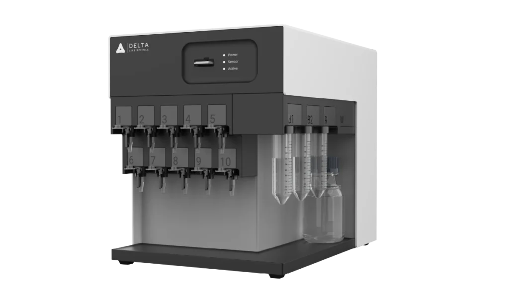
Four independently addressable flow channels, each carrying four sensors, with the sample flowing through the channels in series.
Membrane proteins need a sensor surface built for them, either compatible with a lipid mono- or bilayer, or able to capture a nanodisc with the receptor still functional. A standard hydrogel surface does not do this.
Expression yields for membrane proteins are usually low, so the binding response is small. Baseline noise sits at 0.1 RU RMS, and the sensor is regenerated between injections rather than replaced.
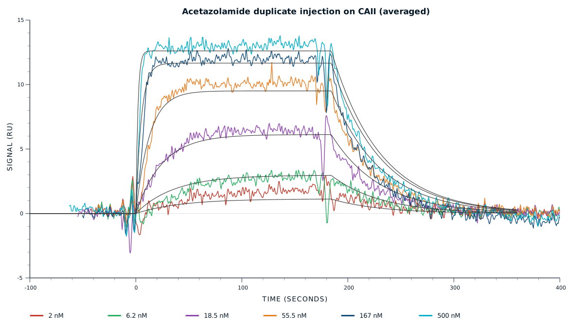
The association phase gives kon, the dissociation phase gives koff, and together they give KD. Affinity runs from 10 pM to 10 mM, with steady-state fitting where dissociation is too fast for kinetics.
Every measurement on a lipid bilayer sensor follows the same steps. The receptor is immobilised in a lipid bilayer on the sensor, the ligand flows past, and the response is recorded through the whole injection.
The receptor is delivered into a bilayer formed across the sensor.
For GPCR and membrane protein binding kinetics analysis, proteoliposomes fuse on the chip to leave a continuous supported bilayer over the waveguide, with the receptor sitting in it in native orientation.
Bare patches left in the layer are blocked before the first ligand sample arrives.
Defects expose the surface underneath, and detergent, free lipid, and buffer components adsorb there readily. That appears in the sensorgram as non-specific binding rather than as receptor engagement.
The ligand flows across the surface and the response climbs.
The association phase gives the on-rate, kon, for receptor-ligand binding. A reference channel carrying a protein-free bilayer of the same lipid composition separates ligand binding from binding to the membrane itself.
Buffer replaces the ligand and the response falls as the complex comes apart.
The dissociation phase gives koff, and 1/koff gives residence time, the number that separates two GPCR ligands sharing a KD. Single-cycle kinetics runs the series uninterrupted when the preparation is scarce.
A mild regeneration buffer returns the sensor to baseline for the next ligand.
The bilayer and the receptor in it stay intact, so every ligand meets the same surface. Conditions are kept gentle, since the lipid environment is what makes the measurement worth having.
Six capabilities, one modular benchtop instrument, in a label-free workflow.
Four channels with in-flow immobilisation, or sixteen sensors with a spotter, compare constructs and controls from one injected sample.
Baseline noise stays low enough that a small binding response is still readable.
One affinity range covers weak modulators through tight ligands, with kon and koff from the same injection series.
Sensor surfaces compatible with lipid mono- and bilayers, or able to capture nanodiscs, keep the receptor functional.
The temperature-controlled flow cell holds conditions steady while a fragile preparation is measured over many injections.
Simply add an autosampler, and run a long construct or condition series unattended.
Capture the receptor on a lipid-compatible sensor surface, or capture the nanodisc carrying it, then inject the ligand across a concentration series. The association phase gives kon, the dissociation phase gives koff, and the two together give KD. Affinity on the inQuiQ runs from 10 pM to 10 mM.
Keep it in a lipid environment. Detergent micelles, nanodiscs, and SMALPs all preserve the folded receptor, and the sensor surface has to suit them. A standard hydrogel chip does not, so membrane work uses a surface built for lipid mono- and bilayers or for nanodisc capture.
Yes, on a sensor surface designed for it. The particle is captured through a tag on the receptor or on the scaffold, and the ligand is then injected over it. A reference channel carrying empty particles separates binding to the receptor from binding to the lipid or the scaffold.
The inQuiQ runs from 30 µL of sample per injection. Sensors are regenerable, so one prepared surface carries a series of ligands, concentrations, and controls rather than one measurement per surface. That matters when expression yield is the limit on the experiment.
Affinity (KD) says how tightly ligand and receptor bind at equilibrium. Kinetics says how they get there: kon for how fast the complex forms, koff for how fast it falls apart. Two ligands with the same KD can have very different residence times.