Targeting-ligand engagement
Confirm the particle reaches the receptor it was designed for.
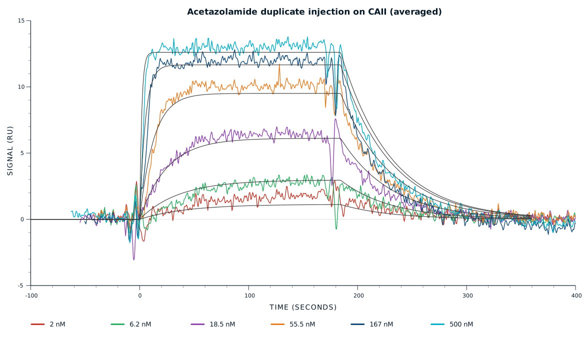
Measure kon and koff at the particle surface.
receptor engagement

Label-free interaction analysis measures binding between unmodified molecules in real time. For EVs and LNPs it returns receptor engagement at the particle surface, association and dissociation rates, and specificity, with no dye or reporter added to the particle.
Confirm the particle reaches the receptor it was designed for.
Measure kon and koff at the particle surface.
Characterise ionizable lipid formulations carrying nucleic acid cargo.
Compare formulations on receptor binding, alongside particle size.
Profile exosome and microvesicle surface proteins.
Screen tetraspanins and other markers against one preparation.
Follow what the particle picks up from its environment.
Measure serum protein binding, ApoE included, at the particle surface.
Separate on-target engagement from surface sticking.
Run competitive and blocking injections against control receptors.
Discuss your assay with one of our application specialists.

Four independently addressable flow channels, each carrying four sensors, with the sample flowing through the channels in series.
The immobilised receptor captures the intact particle, measuring target engagement within the 100 nm sensing area at the sensor surface, not a lysate or released component.
EVs and LNPs diffuse slowly compared with a protein. Flow runs from 5 to 100 µL/min, up to 300 µL/min for low-viscosity samples, so contact time is set to the particle rather than to the instrument.
Compare particle binding to an open target receptor, a blocked receptor, and control surfaces. A strong signal that drops when the receptor is blocked confirms specific engagement.
Every measurement follows the same steps, regardless of the type of particle. The ligand is immobilised on the sensor, the sample flows past, and the response is recorded through the whole injection.
The target receptor goes on the sensor surface first.
For lipid nanoparticle and extracellular vesicle characterisation the receptor, antibody, or surface marker is immobilised and the particle is injected over it. Density is kept low so large particles are not crowded on the surface.
Unused activated sites are switched off before the first particle sample arrives.
Lipid surfaces bind readily to unblocked chemistry. Deactivation, together with the antifouling hydrogel, keeps the sensorgram to receptor engagement rather than particle sticking.
The particle preparation flows across the surface and the response climbs.
In EV and LNP binding kinetics analysis the association phase gives kon for targeting-ligand engagement at the particle surface. Flow rate is lowered so slow-diffusing particles get enough contact time to reach the receptor.
Buffer replaces the particle sample and the response falls as the complex comes apart.
The dissociation phase gives koff, and the pair gives KD for the particle and receptor. Competitive injections with free ligand then separate specific receptor engagement from cross-reactivity.
A regeneration buffer returns the sensor to baseline for the next preparation.
The receptor stays in place, so every LNP formulation or EV batch meets the same surface across a characterisation series.
Six capabilities, one modular benchtop instrument, in a label-free workflow.
Four channels with in-flow immobilisation, or sixteen sensors with a spotter, put target receptors, control receptors, and blanks under the same injected sample.
Baseline noise stays low enough that engagement by a heterogeneous particle population is still readable.
One affinity range covers loose corona interactions and high-affinity targeting-ligand engagement.
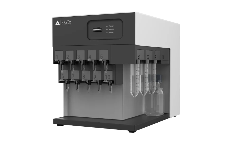
EVs and LNPs diffuse slowly compared with a protein, and flow rate is what decides how long they meet the surface.
Lipid composition and ionic strength differ between formulations, and the same sensor surface carries the comparison across them.
Simply add an autosampler, and run a long formulation or batch series unattended.
Immobilise the target receptor and inject the LNP preparation over it. The association phase gives kon for targeting-ligand engagement, the dissociation phase gives koff, and the two give KD. Nothing is added to the particle, so the surface being measured is the one you formulated.
Spot capture antibodies against tetraspanins and other markers on one chip, then inject the vesicle preparation once. Every sensor reads the same injected sample in series, so a marker profile comes from a single run rather than one assay per marker.
Immobilise the serum protein of interest and inject the particle preparation, or run it the other way round. Real-time recording shows how quickly corona proteins associate and how long they stay, which an endpoint measurement of the settled corona cannot show.
EVs and LNPs diffuse far more slowly than a protein, so at high flow many particles pass without reaching the surface. Flow runs from 5 to 100 µL/min, up to 300 µL/min for low-viscosity samples, which lets contact time be set to the particle rather than to the instrument.
Run the injection over the target receptor, over a control receptor, and over a blank surface, then repeat with the receptor pre-blocked using free ligand. Binding that disappears when the receptor is blocked is receptor engagement; binding that stays is surface sticking.